The 5'->3' Exoribonuclease (XRN) Project
The 5'->3' exoribonucleases (XRNs) comprise a large family of conserved enzymes in eukaryotes with important functions in RNA metabolism and RNA interference, including quality control, degradation, transport, and maturation of ribosomal and small nucleolar RNAs.
XRN1 (175 kD) is primarily cytosolic and is involved in degradation of decapped mRNAs, nonsense mediated decay, microRNA decay and is essential for proper development.
XRN2 (115 kD) is primarily nuclear, and its ortholog in S. cerevisiae, more commonly known as Rat1, was initially shown to have roles in RNA trafficking (ribonucleic acid trafficking 1) from the nucleus to the cytoplasm. More recent studies have identified XRN2/Rat1 as the exoribonuclease that is essential for the 'torpedo' model of transcription termination by RNA polymerase II (Pol II).
The XRNs are found in most eukaryotes, and share two highly conserved regions in their sequences.
XRNs have processive exoribonuclease (with no endoribonuclease) activity towards RNA substrates with a 5' monophosphate group. In comparison, the activity towards RNAs with a 5' hydroxyl, triphosphate or cap is much lower. Both enzymes require divalent cations (Mg2+ or Mn2+) for activity.
The exoribonuclease activity of Rat1 is stimulated by Rai1. Strong sequence homologs of Rai1 are found in other fungal species. A weak sequence homolog of Rai1, known as Dom3Z, exists in mammals, although it does not associate with XRN2. The Rai1 proteins share no recognizable amino acid sequence homology with other proteins.
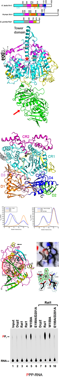
Major findings from this project- The crystal structures of the S. pombe Rat1-Rai1 complex and K. lactis Xrn1 have been determined.
- The two conserved regions of Rat1 and Xrn1 (and XRNs in general) constitute a single, large domain. The active site is formed primarily by residues in the first conserved region. The second conserved region encircles the active site region, consistent with the exclusive exoribonuclease activity of these enzymes.
- The 510-residue segment following the conserved regions in Xrn1 forms four distinct domains. They are located far from the active site but are important for catalysis, possibly by stabilizing the N-terminus of the enzyme.
- Helix aD in the first conserved region of Rat1 is exceptionally long (with 35 residues), and its C-terminal end is projected 30A away from the rest of the structure. We have named this feature of the Rat1 structure the 'tower' domain. This domain may be unique to XRN2 and may serve XRN2-specific functions.
- Rai1 is bound near the bottom of Rat1, about 30A away and on the opposite face of the protein from the active site. Mutations of residues in the interface can abolish the interactions between the two proteins.
- Our structural data, together with earlier biochemical data, demonstrate that the activating effect of Rai1 is due to its stabilization of the Rat1 structure.
- The structures of Rai1 and Dom3Z do not appear to have a close homolog in the Protein Data Bank, and they may represent a new protein fold.
- There is a large pocket in the surfaces of Rai1 and Dom3Z, and many of the side chains in this pocket are highly conserved among Rai1 homologs.
- Most importantly, there is a cluster of four acidic residues (Glu150, Glu199, Asp201, and Glu239 in Rai1) at the bottom of the pocket, and three of them (Glu150, Asp201, and Glu239), together with the main chain carbonyl of Leu240 and two water molecules, form the octahedral coordination sphere of a divalent cation (Mg2+ or Mn2+).
- The structural information therefore suggests that Rai1 and Dom3Z may have catalytic functions. This putative active site is distinct from the Rat1-Rai1 interface.
- Biochemical studies confirm that Rai1 does possess catalytic activity, acting as a pyrophosphohydrolase on RNA substrates with a 5' triphosphate.
- Rai1 and Dom3Z may have important roles in mRNA quality surveillance, promoting the degradation of mRNAs with defective 5' caps.
Publications from this project
- Xiang S, Cooper-Morgan A, Jiao X, Kiledjian M, Manley JL, Tong L. (2009). Structure and function of the 5'->3' exoribonuclease Rat1 and its activating partner Rai1. Nature, 458, 784-788. Reprint(PDF)
- Chang J.H., Xiang S, Xiang K, Manley JL, Tong L. (2011). Structural and biochemical studies of the 5'->3' exoribonuclease Xrn1. Nature Struct. Mol. Biol., 18, 270-276. Reprint(PDF)
- Chang J.H., Xiang S, Tong L. (2011). 5'->3' exoribonucleases. Ribonucleases, Chapter 7, pp. 167-192. A.W. Nicholson, Ed. Nucleic Acids and Molecular Biology 26, Springer-Verlag. Reprint(PDF)
Funding for this project
- NIH GM090059
© copyright 2009-2018, Liang Tong.